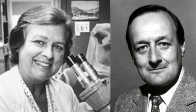
 Image 5: Dr. Margaret Billingham (left) and Dr. Philip Caves ( right)
Endomyocardial Biopsy
After heart transplants were a success, the attention turned to preventing the patient's immune system from rejecting the new organ. To do this, Dr. Margaret Billingham and Dr. Philip Caves developed a procedure that used endomyocardial biopsy as a method to view tissue rejection. Endomyocardial biopsy is the removal of the heart's inner tissue. To do this, they would insert a thin instrument down the patient's neck vein and into the heart's chamber where they would gather pieces of myocardial tissue under fluoroscopic control.Through this procedure they were able to determine based on Dr. Billingham's grading rejection scale if they needed to give the patient stronger doses of anti-rejection medication to save the organ. This method decreased the number of deaths of patients with heart transplants due to organ rejection, in fact, it was so successful the grading system is still used today. The scale consisted of four levels: OR , 1R mild, 2R moderate, and 3R severe. OR meant there was no evidence of organ rejection, heart tissue was healthy; 1R showed one focus of myocyte (muscle cell) damage; 2R showed two or more foci of myocyte damage and lastly, 3R was the most severe case broadcasting multifocal myocyte damage, edema ( swelling), hemorrhage ( profuse bleeding) and vasculitis (inflammation of the blood vessels).
After heart transplants were a success, the attention turned to preventing the patient's immune system from rejecting the new organ. To do this, Dr. Margaret Billingham and Dr. Philip Caves developed a procedure that used endomyocardial biopsy as a method to view tissue rejection. Endomyocardial biopsy is the removal of the heart's inner tissue. To do this, they would insert a thin instrument down the patient's neck vein and into the heart's chamber where they would gather pieces of myocardial tissue under fluoroscopic control.Through this procedure they were able to determine based on Dr. Billingham's grading rejection scale if they needed to give the patient stronger doses of anti-rejection medication to save the organ. This method decreased the number of deaths of patients with heart transplants due to organ rejection, in fact, it was so successful the grading system is still used today. The scale consisted of four levels: OR , 1R mild, 2R moderate, and 3R severe. OR meant there was no evidence of organ rejection, heart tissue was healthy; 1R showed one focus of myocyte (muscle cell) damage; 2R showed two or more foci of myocyte damage and lastly, 3R was the most severe case broadcasting multifocal myocyte damage, edema ( swelling), hemorrhage ( profuse bleeding) and vasculitis (inflammation of the blood vessels).
